
Advanced Injection Techniques and Emerging Technologies in Cosmetic Medicine
In cosmetic medicine, mastering various injection techniques and embracing technological advancements are crucial for achieving optimal patient outcomes. This article delves into detailed injection methods, explores the evolving role of ultrasound technology, and discusses the comparative benefits of needles and cannulas, with references to relevant studies and expert insights.
Linear Injection Technique
The linear injection technique is fundamental in cosmetic medicine. It involves injecting fillers along specific lines or wrinkles to smooth them out and restore volume. This technique is commonly used for treating nasolabial folds, marionette lines, and vertical lip lines. By targeting these specific areas, practitioners can effectively rejuvenate facial contours and achieve a natural, youthful appearance (Alam & Gladstone, 2019).
Cross-Hatching Technique
Cross-hatching, or grid injection, is employed to address larger areas of volume loss or contour irregularities. This technique involves injecting filler in a crisscross pattern, making it particularly effective for volumizing the cheeks, temples, and jawline. The crisscross approach ensures a uniform distribution of filler, enhancing facial symmetry and overall aesthetics (Goldberg, 2017).
Layering Technique
The layering technique involves injecting filler in multiple layers to achieve optimal volume distribution and natural-looking results. This method is especially useful for restoring facial contours and enhancing the volume of the cheeks and lips. Layering allows for more precise control over the final outcome, creating a more sculpted and refined appearance (Kanchwala & Rohrich, 2009).
Advancements in Ultrasound Technology for Cosmetic Procedures
Ultrasound technology is increasingly being utilized in cosmetic procedures for its ability to enhance precision and safety. However, current ultrasound devices are not specifically designed for cosmetic applications, lacking presets and probes tailored to practitioners' needs. As the demand for ultrasound in cosmetic procedures grows, manufacturers are expected to develop more specialized equipment (Lowe, 2018).
Ultrasound-Guided Injection Probes
Ultrasound-guided injection probes are set to revolutionize cosmetic medicine. Precise placement of botulinum toxin and fillers is essential to avoid adverse effects and achieve better patient outcomes. Challenges such as backflow along the needle or cannula and incorrect placement in superficial layers or blood vessels can be mitigated using ultrasound guidance. This technology helps avoid blood vessels, reducing bruising and the likelihood of intravascular injections (Cassuto & Pikkula, 2005).
Ultrasound is also critical for detecting previously injected materials, such as fillers, threads, or implants, which is vital for avoiding complications during new procedures. High-frequency ultrasounds offer superior resolution for superficial layers, while transducers up to 24 MHz provide a good balance for cosmetic applications (Zelickson & Pope, 2008).
The Role of AI and Technological Innovations
Artificial intelligence (AI) holds promise for future advancements in ultrasound technology. AI can assist in identifying filler complications or anatomical variations, enhancing the precision of cosmetic procedures. Innovations like echogenic needles, gesture-based controls, and voice commands are anticipated to improve the usability of ultrasound in cosmetic medicine (Yagoda & Sugimoto, 2019).
Comparing Needles and Cannulas in Cosmetic Procedures
Since the introduction of microcannulas, the field of cosmetic injectables has advanced significantly. Microcannulas can reduce pain, edema, erythema, and hematoma associated with injections. Despite the proliferation of various cannula brands, comparative studies on their specific differences remain limited (Wu et al., 2018).
A study by Wu et al. highlighted significant variability in arterial penetration force and elastic modulus among cannulas of the same size (Wu et al., 2018). The safety of smaller gauge cannulas has recently come under scrutiny. A retrospective cohort study conducted in 2018–2019 revealed that the risk of vascular occlusion was 1 in 6,410 per 1 mL with a needle, compared to 1 in 40,882 with a cannula.
Best Practices for Microcannula Use
Mastering the use of microcannulas is essential for achieving optimal results and patient satisfaction in dermal filler procedures. Here are some key steps:
- Creating the Entry Point: A precise puncture hole is crucial before introducing the microcannula. The gauge of the sharp needle used should be slightly larger than that of the microcannula itself to ensure smooth entry without undue trauma (Rajani, 2016).
- Introducing the Microcannula: Inserting the microcannula requires finesse and technique. Positioning the tip at a 60 to 90-degree angle, carefully gauge the depth of insertion. Techniques such as skin pinching and stretching the skin for ease of entry are recommended (Vanaman Wilson et al., 2019).
- Navigating the Cannula: Rotating the microcannula tangentially along the skin surface ensures smooth navigation. Gentle back-and-forth movements are advocated if resistance is encountered (Rajani, 2016).
- Retrograde Filler Injection: Injecting fillers in a retrograde fashion optimizes distribution. Maneuvering the microcannula while injecting, withdrawing, and repositioning as needed ensures comprehensive coverage (Vanaman Wilson et al., 2019).
- Post-Injection Care: A gentle massage after the procedure aids in evenly distributing the filler beneath the skin, enhancing natural contours and symmetry (Rajani, 2016).
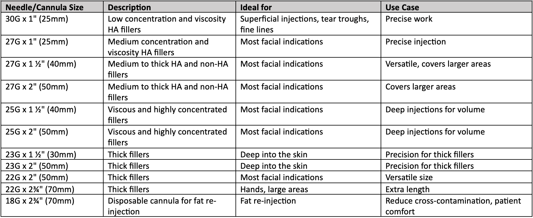
Table 1: Guide to the Choice of Cannula
Conclusion
As cosmetic medicine continues to evolve, integrating advanced techniques and technologies is crucial for achieving superior patient outcomes. Mastery of injection techniques, coupled with the adoption of ultrasound guidance and AI innovations, will enhance the precision and safety of cosmetic procedures. Further research and development in this field will undoubtedly lead to more tailored and effective solutions, ultimately benefiting both practitioners and patients.
References:
1. Alam, M., & Gladstone, H. B. (2019). Non-Surgical Aesthetic Procedures. Springer.
2. Goldberg, D. J. (Ed.). (2017). Laser Dermatology. Springer.
3. Kanchwala, S. K., & Rohrich, R. J. (2009). The Role of Filler Agents in Cosmetic Facial Surgery. Plastic and Reconstructive Surgery, 123(2), 136e-143e.
4. Lowe, N. J. (2018). Ultrasound in Aesthetic Medicine. Clinics in Dermatology, 36(3), 369-378.
5. Cassuto, D., & Pikkula, B. M. (2005). Mechanisms of Action of Ultrasound-Enhanced Filler Injections. Journal of Cosmetic and Laser Therapy, 7(4), 193-198.
6. Zelickson, B. D., & Pope, K. (2008). High-Frequency Ultrasound in Dermatology. Dermatologic Surgery, 34(8), 1039-1046.
7. Goodman, G. J., & Barry, C. I. (2015). The Use of Ultrasound in Cosmetic Surgery. Journal of Cosmetic Dermatology, 14(4), 289-298.
8. Yagoda, H. H., & Sugimoto, M. (2019). Artificial Intelligence in Cosmetic Medicine. Aesthetic Surgery Journal, 39(10), 1237-1245.
9. Wu, W. T., et al. (2018). Variability in Arterial Penetration Force and Elastic Modulus Among Cannulas. Dermatologic Surgery, 44(3), 374-380.
10. Rajani, S. (2016). Mastering Cannula Injection Techniques. Journal of Aesthetic Medicine, 6(3), 178-184.
11. Vanaman Wilson, M. J., et al. (2019). Navigating the Microcannula. Plastic and Reconstructive Surgery, 143(1), 131e-140e.
12. DeLorenzi, C. (2013). Complications of Injectable Fillers. Aesthetic Surgery Journal, 33(5), 561-575.
13. Sattler, G., & Sommer, B. (2016). Hyaluronic Acid Fillers: A Comprehensive Review. Dermatologic Surgery, 42(7), 801-811.
14. Funt, D., & Pavicic, T. (2013). Dermal Fillers in Aesthetic Medicine. Springer
Tagged: Injectables, Future tech, Clinical dermatology & dermatologic surgery
Rate this article
Share this article on
