
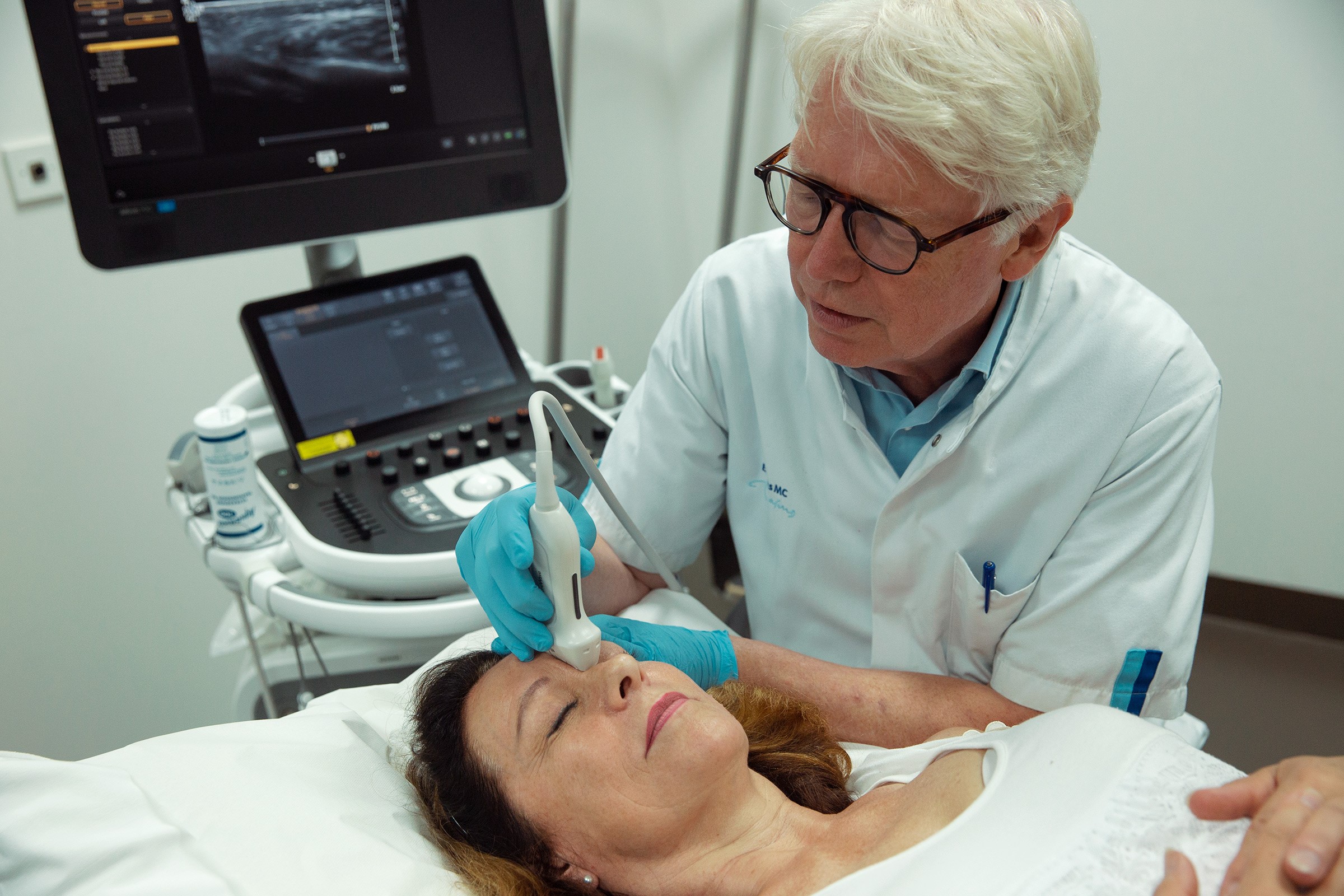
Dr Peter Velthuis talks about the growing importance of ultrasonography for injections
The 2022 edition of the IMCAS World Congress takes place in person (finally!) in Paris from January 27 to 29. A key scientific highlight this year is on the topic of ultrasonography and its growing importance when it comes to injectables and thread treatments. To introduce this better, IMCAS reached out to a top expert in the subject of ultrasonography.
Meet Dr Peter Velthuis, our go-to ultrasound-master who heads the Cutaneous group , a scientific partner of IMCAS who offers professional courses on facial ultrasound. You can see him in person at the IMCAS World Congress 2022 during sessions dedicated to clinical dermatology, lasers, complications & ultrasonography.
1. What is the role of ultrasound in aesthetic practices? What does it show and how is it used?
The most exiting application of ultrasound in filler medicine is seeing the injection material being delivered in real time. When you observe that for the first time it is a real whoo-effect. But in general physicians tend to use it for prevention. So, prior to injecting in a certain area they inspect that with duplex to see if there are any arteries in the way. Checking your treatment is another important use. You are able to detect exactly the location the filler is injected. That is sometimes confronting, but it really helps you to improve your technique. Then of course, we have been using is for over 10 years to treat complications. Finally, ultrasound imaging of the face will enable you to learn more about anatomy.
2. What is a typical example of complication that could have been avoided with ultrasound?
Most important is to stay away from arteries to avoid intravascular injection. But to assess an area anatomically can also help. For instance, we see many filler injections inside the parotid gland. Measuring the subcutaneous space in the area makes you aware that the margins are thin, sometimes only 2 millimeters.
3. How did you begin to delve into this subject?
As a dermatologist I was also trained as phlebologist. Picking up the ultrasound probe when I had my first filler complications seemed as a natural thing to do. This was in 2011. Together with Dr Leonie Schelke, I became part of the dermatology department of Erasmus University Medical Center in Rotterdam. However, it took us years to understand that ultrasound imaging could give much more information that just how the filler was looking. And being at a university our task was to investigate. So that is how we started to publish scientific papers.
Now it is our aim to have all doctors realize the importance of ultrasound imaging of the face for filler injections. It takes an effort to learn this stuff. But it is very rewarding and I am convinced that in a few years’ time all clients are going to expect his/her doctor to use a device prior to injecting.
4. Is ultrasound easy to incorporate into aesthetic practice? Or does it take some training before hand?
It is not easy to start with ultrasound in your practice. You have to be committed to do so. If you are, you need to buy a device and read about this subject. But reading only is not enough. It really helps to take classes to delve deeper, like we are doing during IMCAS World Congress 2022 where there will be a special highlight on ultrasonography through sessions and exhibitions. You also need hands-on training. The upcoming AATC in January 2021 in Paris, the two days before IMCAS would be a great introduction. Then after you have mastered the basics, it is like learning to drive a car. Practice, practice, practice…
Make sure the device is in reaching distance with every client that you see. And even when you think you do not see enough, your clients will love you for examining them.
Many doctors charge a little extra for this service and people do not mind to pay for that, we found out.
Mots-clés: Dermatologie clinique & chirurgie dermatologique
Noter cet article
Partagez cet article sur